Probing electrical signals in the retina via graphene-integrated microfluidic platforms
Abstract
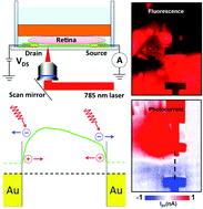
Graphene has attracted extensive attention in biological and biomedical fields due to its unique physical properties and excellent biocompatibility. We combine graphene field-effect transistors and scanning photocurrent microscopy with microfluidic platforms to investigate electrical signals in mouse retina. Remarkable photocurrent signals were detected from the graphene underneath the optic nerve head (ONH) of the retina, where the electrical activity from this region can modulate the carrier concentration of the graphene and induce local potential gradients. These built-in electrical potential gradients can efficiently separate photo-excited electron–hole pairs, leading to strong photocurrent responses in the graphene underneath the ONH. We also show that no significant photocurrent signal was observed in the graphene underneath either dehydrated or fixed retinal tissues, verifying that the photocurrent responses generated in the graphene underneath the ONH were indeed induced by the electrical activity in living retina. This method not only provides a way to investigate electrical processes in living retinal tissues, but also offers opportunities to study many other cellular systems involving cell–cell interactions through electrical signaling.
 Please wait while we load your content...

