Manganese-impregnated mesoporous silica nanoparticles for signal enhancement in MRI cell labelling studies†
Abstract
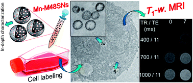
Mesoporous silica nanoparticles (MSNs) are used in drug delivery and cell tracking applications. As Mn2+ is already implemented as a “positive” cell contrast agent in preclinical imaging procedures (in the form of MnCl2 for neurological studies), the introduction of Mn in the porous network of MSNs would allow labelling cells and tracking them using MRI. These particles are in general internalized in endosomes, an acidic environment with high saline concentration. In addition, the available MSN porosity could also serve as a carrier to deliver medical/therapeutic substances through the labelled cells. In the present study, manganese oxide was introduced in the porous network of MCM-48 silica nanoparticles (Mn–M48SNs). The particles exhibit a narrow size distribution (∼140 nm diam.) and high porosity (∼60% vol.), which was validated after insertion of Mn. The resulting Mn–M48SNs were characterized by TEM, N2 physisorption, and XRD. Evidence was found with H2-TPR, and XPS characterization, that Mn(II) is the main oxidation state of the paramagnetic species after suspension in water, most probably in the form of Mn–OOH. The colloidal stability as a function of time was confirmed by DLS in water, acetate buffer and cell culture medium. In NMR data, no significant evidence of Mn2+ leaching was found in Mn–M48SNs in acidic water (pH 6), up to 96 hours after suspension. High longitudinal relaxivity values of r1 = 8.4 mM−1 s−1 were measured at 60 MHz and 37 °C, with the lowest relaxometric ratios (r2/r1 = 2) reported to date for a Mn–MSN system. Leukaemia cells (P388) were labelled with Mn–M48SNs and nanoparticle cell internalization was confirmed by TEM. Finally, MRI contrast enhancement provided by cell labelling with escalated incubation concentrations of Mn–M48SNs was quantified at 1 T. This study confirmed the possibility of efficiently confining Mn into M48SNs using incipient wetness, while maintaining an open porosity and relatively high pore volume. Because these Mn-labelled M48SNs express strong “positive” contrast media properties at low concentrations, they are potentially applicable for cell tracking and drug delivery methodologies.
- This article is part of the themed collection: Functional Nanoparticles for Biomedical Applications
 Please wait while we load your content...

