Organic-to-water dispersible Mn:ZnS–ZnS doped core–shell quantum dots: synthesis, characterization and their application towards optical bioimaging and a turn-off fluorosensor†

Abstract
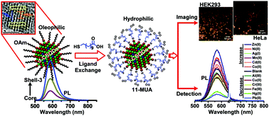
In the present study, core–shell quantum dots (CSQDs) containing environmentally-benign transition metal ion Mn(II) doped ZnS (Mn:ZnS) as a core material encapsulated within different thickness ZnS shell layers were synthesized and studied. The Mn:ZnS core was synthesized by a heating-up method, and a hot-injection technique was used to encapsulate the core within ZnS shell layers of different thicknesses. The doped core–shell quantum dots (d-CSQDs) in a dot-in-dot architecture possess polytypism in zinc blende (core)/wurtzite (shell) crystalline phases. The resultant core d-QDs have a narrow size distribution with a mean diameter of 2.8 nm and it was increased to 4.4 nm after overcoating the ZnS shell layers. The d-CSQDs exhibited a tailored optical bandgap ranging from 3.63 to 3.90 eV, and well-resolved Mn2+ spin-flip emission with a maximum net improved quantum yield up to 38.6% and longer phosphorescence lifetime up to ∼2.67 milliseconds, which signified their excellent optical properties. The mechanism for various emissions emerging in d-QDs is well articulated. In order to evaluate the capability of Mn:ZnS–ZnS d-CSQDs as a fluorescent probe, preliminary experiments have been employed to switch the hydrophobic d-CSQDs to hydrophilic ones by exchanging the native cap with 11-mercaptoundecanoic acid. The resultant colloidal hydrophilized d-CSQDs have shown robust photostability under continuous UV-irradiation for 24 h. These photostable hydrophilized d-CSQDs were further scrutinized in terms of cell viability and cellular internalization. Two different cell lines have been used as testing cells, namely HEK-293 (human embryonic kidney cell line) and HeLa (cervical cancer cell line). The results highlight the capability of these colloidal d-CSQDs to optically image cells without being destructive towards them. Furthermore, it was demonstrated that hydrophilized d-CSQDs can be used as a sensitive “turn-off” fluorosensor for the detection of Hg2+ and Pb2+ cations with a lower detection limit of 16.3 nM and 8.0 nM respectively.
 Please wait while we load your content...

