
Fabrication of an l-glutathione sensor based on PEG-conjugated functionalized CNT nanocomposites: a real sample analysis
 *ab
*ab
Abstract
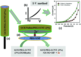
Three series of polyethylene glycol–carbon nanotube nanocomposites in the form of PEG/CNTa–e, PEG/f-CNT.Oxia–e, and PEG/CNT.C18a–e have been fabricated using a dissolution stirring ultra-sonication method. The compounds were then characterized using different characterization techniques, including FT-IR, XRD, and TEM. The FT-IR and XRD data provided clear evidence of the CNTs functionalization as well as the composite formation. The FT-IR spectral data of the functionalized MWCNTs were slightly changed due to the introduction of PEG groups on their surfaces. The TEM images indicated that the nanotubes were homogenously distributed and embedded within the PEG polymer matrix. A flat glassy carbon electrode was modified with GCE/PEG-f-CNToxd nanocomposites (NCs) to obtain a sensor for L-glutathione (GSH), which was seen to exhibit improved sensitivity, low detection limit, large dynamic range, short response time, and good stability, whereby the calibration plot (at +0.5 V) was linear (r2: 0.9957) in the 0.1 nM to 1.0 mM GSH concentration range, the detection limit was as low as 0.026 nM, and the sensitivity was 17.4 μA μM−1 cm−2. To the best of our knowledge, this is the first report on the determination of GSH using such a modified GCE/PEG-f-CNTs by an I–V approach. The GCE/PEG-f-CNT was applied for the selective determination of GSH in spiked rabbit serum samples and achieved acceptable results.
 Please wait while we load your content...

