Issue 6, 2019

Indirect magnetic force microscopy

Abstract

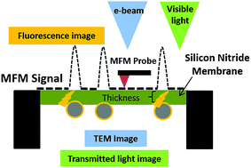
Magnetic force microscopy (MFM) is an atomic force microscopy (AFM)-based technique to map magnetic domains in a sample. MFM is widely used to characterize magnetic recording media, magnetic domain walls in materials, nanoparticles and more recently iron deposits in biological samples. However, conventional MFM requires multiple scans of the samples, suffers from various artifacts and is limited in its capability for multimodal imaging or imaging in a fluid environment. We propose a new modality, namely indirect magnetic force microscopy (ID-MFM), a technique that employs an ultrathin barrier between the probe and the sample. Using fluorescently conjugated superparamagnetic nanoparticles, we demonstrate how ID-MFM can be achieved using commercially available silicon nitride windows, MFM probes and AFM equipment. The MFM signals obtained using ID-MFM were comparable to those obtained using conventional MFM. Further, samples prepared for ID-MFM were compatible with multi-modal imaging via fluorescence and transmission electron microscopy. Thus ID-MFM can serve as a high-throughput, multi-modal microscopy technique which can be especially attractive for detecting magnetism in nanoparticles and biological samples.

Graphical abstract: Indirect magnetic force microscopy

Supplementary files

Article information

Article type
Paper
Submitted
29 Mar 2019
Accepted
03 May 2019
First published
08 May 2019
This article is Open Access
Creative Commons BY-NC license

Nanoscale Adv., 2019,1, 2348-2355

Indirect magnetic force microscopy

J. Sifford, K. J. Walsh, S. Tong, G. Bao and G. Agarwal, Nanoscale Adv., 2019, 1, 2348 DOI: 10.1039/C9NA00193J

This article is licensed under a Creative Commons Attribution-NonCommercial 3.0 Unported Licence. You can use material from this article in other publications, without requesting further permission from the RSC, provided that the correct acknowledgement is given and it is not used for commercial purposes.

To request permission to reproduce material from this article in a commercial publication, please go to the Copyright Clearance Center request page.

If you are an author contributing to an RSC publication, you do not need to request permission provided correct acknowledgement is given.

If you are the author of this article, you do not need to request permission to reproduce figures and diagrams provided correct acknowledgement is given. If you want to reproduce the whole article in a third-party commercial publication (excluding your thesis/dissertation for which permission is not required) please go to the Copyright Clearance Center request page.

Read more about how to correctly acknowledge RSC content.

Social activity

Spotlight

Advertisements