
Particle size-independent induction of leucism in Drosophila melanogaster by silver: nano vs. micro†
Abstract
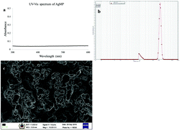
With the discovery of nanoscience, silver nanoparticles (AgNPs) now appear on the ingredient lists of commercial products along with bulk silver or silver microparticles (AgMPs). In the present study, we have compared the effects of silver in both nano and micro forms to assess whether particle size plays a role in the pigmentation pathway, using Drosophila melanogaster as a model organism. AgNPs were synthesized, characterized and validated. Internalization of the AgNPs and AgMPs was confirmed by atomic absorption spectroscopy. Analysis of phenol oxidase (PO) enzyme and total melanin as well as quantification of tyrosine and dihydroxy phenylalanine (dopa) were carried out to understand the perturbation of the melanization pathway. The interactions of AgNP/MP with tyrosine and dopa were investigated using various spectrometric techniques. The absence of PO activity, reduced levels of melanin, tyrosine, and dopa, and the absence of local dissemination of melanin upon cuticle injury confirmed that the AgNPs and AgMPs mediated leucism in Drosophila. The reduction of bulk silver to the nano form upon internalization was found to be the modus operandi for the AgMPs. The actions of the AgNPs and AgMPs were attributed to their binding and interaction with tyrosine and dopa via their phenolic hydroxyl groups. Silver, irrespective of its size, induced leucism in Drosophila melanogaster by binding and interacting with precursor metabolites of the melanization pathway, such as tyrosine and dopa.
 Please wait while we load your content...