
Quantitative elemental analysis of bovine ovarian follicles using X-ray fluorescence imaging
Abstract
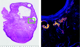
The X-ray Fluorescence Micro-spectroscopy (XFM) beamline at the Australian Synchrotron was used to image 97 follicle histological sections from 45 different bovine ovaries focusing on healthy antral follicles ranging from small (<4 mm) up to preovulatory sizes (>16 mm) and on antral follicles undergoing atresia. This analysis identified five elements (Cu, Fe, Zn, Se and Br) consistently present within the ovarian tissue with Fe, Zn and Se localised to specific structures. GeoPIXE v6.4g was subsequently used to extract quantitative information pertaining to the elemental concentrations surrounding each of these follicles. Statistical analysis suggested that significant elemental differences were evident between follicle groups sorted according to their health status (Fe and Br), and their size (Se). Se appeared to be the element which most greatly distinguished large antral follicles from smaller counterparts. The ability to use synchrotron radiation to measure trace element distributions in bovine follicles at such high resolutions could have a significant impact on understanding the mechanisms of follicular development. This research is intended to form a baseline study of healthy cycling ovaries which could later be extended to disease states, thereby improving our current understanding of infertility and endocrine diseases involving the ovary.
 Please wait while we load your content...