Determination of elemental distribution in green micro-algae using synchrotron radiation nano X-ray fluorescence (SR-nXRF) and electron microscopy techniques – subcellular localization and quantitative imaging of silver and cobalt uptake by Coccomyxa actinabiotis†
Abstract
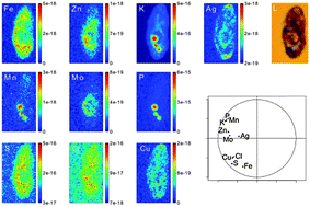
The newly discovered unicellular micro-alga Coccomyxa actinabiotis proves to be highly radio-tolerant and strongly concentrates radionuclides, as well as large amounts of toxic metals. This study helps in the understanding of the mechanisms involved in the accumulation and detoxification of silver and cobalt. Elemental distribution inside Coccomyxa actinabiotis cells was determined using synchrotron nano X-ray fluorescence spectroscopy at the ID22 nano fluorescence imaging beamline of the European Synchrotron Radiation Facility. The high resolution and high sensitivity of this technique enabled the assessment of elemental associations and exclusions in subcellular micro-algae compartments. A quantitative treatment of the scans was implemented to yield absolute concentrations of each endogenous and exogenous element with a spatial resolution of 100 nm and compared to the macroscopic content in cobalt and silver determined using inductively coupled plasma-mass spectrometry. The nano X-ray fluorescence imaging was complemented by transmission electron microscopy coupled to X-ray microanalysis (TEM-EDS), yielding differential silver distribution in the cell wall, cytosol, nucleus, chloroplast and mitochondria with unique resolution. The analysis of endogenous elements in control cells revealed that iron had a unique distribution; zinc, potassium, manganese, molybdenum, and phosphate had their maxima co-localized in the same area; and sulfur, copper and chlorine were almost homogeneously distributed among the whole cell. The subcellular distribution and quantification of cobalt and silver in micro-alga, assessed after controlled exposure to various concentrations, revealed that exogenous metals were mainly sequestered inside the cell rather than on mucilage or the cell wall, with preferential compartmentalization. Cobalt was homogeneously distributed outside of the chloroplast. Silver was localized in the cytosol at low concentration and in the whole cell excluding the nucleus at high concentration. Exposure to low concentrations of cobalt or silver did not alter the localization nor the concentration of endogenous elements within the cells. To our knowledge, this is the first report on element co-localization and segregation at the sub-cellular level in micro-algae by means of synchrotron nano X-ray fluorescence spectroscopy.
- This article is part of the themed collection: Fourth International Symposium on Metallomics, Oviedo, Spain
 Please wait while we load your content...