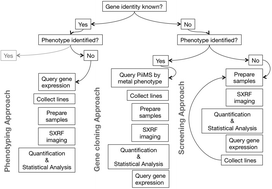
Here we present approaches for using multi-elemental imaging (specifically synchrotron X-ray fluorescence microscopy, SXRF) in ionomics, with examples using the model plant Arabidopsis thaliana. The complexity of each approach depends on the amount of a priori information available for the gene and/or phenotype being studied. Three approaches are outlined, which apply to experimental situations where a gene of interest has been identified but has an unknown phenotype (phenotyping), an unidentified gene is associated with a known phenotype (gene cloning) and finally, a screening approach, where both gene and phenotype are unknown. These approaches make use of open-access, online databases with which plant molecular genetics researchers working in the model plant Arabidopsis will be familiar, in particular the Ionomics Hub and online transcriptomic databases such as the Arabidopsis eFP browser. The approaches and examples we describe are based on the assumption that altering the expression of ion transporters can result in changes in elemental distribution. We provide methodological details on using elemental imaging to aid or accelerate gene functional characterization by narrowing down the search for candidate genes to the tissues in which elemental distributions are altered. We use synchrotron X-ray microprobes as a technique of choice, which can now be used to image all parts of an Arabidopsis plant in a hydrated state. We present elemental images of leaves, stem, root, siliques and germinating hypocotyls.
You have access to this article
 Please wait while we load your content...
Something went wrong. Try again?
Please wait while we load your content...
Something went wrong. Try again?