
Stable iron isotope tracing reveals significant brain iron uptake in adult rats
Abstract
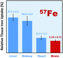
Iron deposits in the brain are a common hallmark of Alzheimer's disease and Parkinson's disease. This has spurred the hypothesis that iron may play a functional role in the pathogenesis of neurodegenerative disorders through free radical damage. Previous short-term studies using radiotracers suggested that brain iron uptake is small as compared to other tissues in adult rodents. This has led to the assumption that brain iron uptake must also be marginal in humans after brain development is complete. In this study we applied a novel approach to determine directly the fraction of iron that was transferred over time from diet to brain and other organs in adult rats. A known amount of a stable iron isotope (57Fe) was fed with drinking water to adult rats over 4 months. Uptake of the tracer iron and final iron content in tissues were assessed by Negative Thermal Ionization Mass Spectrometry (NTI-MS). We found that only a very small amount of dietary iron entered the brain (0.000537 ± 0.000076%). This amount, however, is considerable relative to the total brain iron content (9.19 ± 0.71%), which was lower but comparable to percentage uptake in other tissues. Whereas it remains unclear whether excessive dietary iron intake is a risk factor in neurodegenerative diseases or whether high systemic iron correlates with iron deposits in the brain, our study suggests that uptake of dietary iron is much higher than previously thought. This finding challenges current beliefs and points to a possible role of iron nutrition in the pathogenesis of neurodegenerative disorders.
 Please wait while we load your content...