Microfluidic bead trap as a visual bar for quantitative detection of oligonucleotides†
 *abcd
*abcd
Abstract
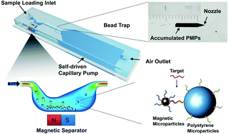
We demonstrate a microfluidic bead trap capable of forming a dipstick-type bar visible to the naked eye for simple and quantitative detection of oligonucleotides. We use magnetic microparticles (MMPs) and polystyrene microparticles (PMPs) that are connected and form MMPs–targets–PMPs when target oligonucleotides are present, leaving free PMPs with a number inversely proportional to the amount of targets. Using a capillary flow-driven microfluidic circuitry consisting of a magnetic separator to remove the MMPs–targets–PMPs, the free PMPs can be trapped at the narrowing nozzle downstream, forming a visual bar quantifiable based on the length of PMP accumulation. Such a power-free and instrument-free platform enables a limit of detection at 13 fmol (0.65 nM in 20 μl, S/N = 3) of oligonucleotides and is compatible with single-nucleotide polymorphisms and operation in a complex bio-fluid. Moreover, using DNAzyme as the target oligonucleotide that catalyzes a specific hydrolytic cleavage in the presence of lead ions, we demonstrate a model application that detects lead ions with a limit of detection of 12.2 nM (2.5 μg l−1), providing quantitative and visual detection of lead contamination at resource-limited sites.
 Please wait while we load your content...

