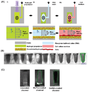
Hydrogel-coated microfluidic channels for cardiomyocyte culture†
Abstract
The research areas of tissue engineering and
- This article is part of the themed collection: Organs on a Chip 2013
The research areas of tissue engineering and
N. Annabi, Š. Selimović, J. P. Acevedo Cox, J. Ribas, M. Afshar Bakooshli, D. Heintze, A. S. Weiss, D. Cropek and A. Khademhosseini, Lab Chip, 2013, 13, 3569 DOI: 10.1039/C3LC50252J
This article is licensed under a Creative Commons Attribution 3.0 Unported Licence. You can use material from this article in other publications without requesting further permissions from the RSC, provided that the correct acknowledgement is given.
Read more about how to correctly acknowledge RSC content.
 Please wait while we load your content...

