Issue 22, 2012

A novel microfluidic platform for high-resolution imaging of a three-dimensional cell culture under a controlled hypoxic environment

Abstract

Low oxygen tensions experienced in various pathological and physiological conditions are a major stimulus for angiogenesis. Hypoxic conditions play a critical role in regulating cellular behaviour including migration, proliferation and differentiation. This study introduces the use of a microfluidic device that allows for the control of oxygen tension for the study of different three-dimensional (3D) cell cultures for various applications. The device has a central 3D gel region acting as an external cellular matrix, flanked by media channels. On each side, there is a peripheral gas channel through which suitable gas mixtures are supplied to establish a uniform oxygen tension or gradient within the device. The effects of various parameters, such as gas and media flow rates, device thickness, and diffusion coefficients of oxygen were examined using numerical simulations to determine the characteristics of the microfluidic device. A polycarbonate (PC) film with a low oxygen diffusion coefficient was embedded in the device in proximity above the channels to prevent oxygen diffusion from the incubator environment into the polydimethylsiloxane (PDMS) device. The oxygen tension in the device was then validated experimentally using a ruthenium-coated (Ru-coated) oxygen-sensing glass cover slip which confirmed the establishment of low uniform oxygen tensions (<3%) or an oxygen gradient across the gel region. To demonstrate the utility of the microfluidic device for cellular experiments under hypoxic conditions, migratory studies of MDA-MB-231 human breast cancer cells were performed. The microfluidic device allowed for imaging cellular migration with high-resolution, exhibiting an enhanced migration in hypoxia in comparison to normoxia. This microfluidic device presents itself as a promising platform for the investigation of cellular behaviour in a 3D gel scaffold under varying hypoxic conditions.
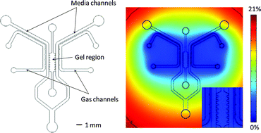
Graphical abstract: A novel microfluidic platform for high-resolution imaging of a three-dimensional cell culture under a controlled hypoxic environment

Supplementary files

Article information

Article type
Paper
Submitted
30 Mar 2012
Accepted
04 Sep 2012
First published
04 Sep 2012

Lab Chip, 2012,12, 4855-4863

A novel microfluidic platform for high-resolution imaging of a three-dimensional cell culture under a controlled hypoxic environment

K. Funamoto, I. K. Zervantonakis, Y. Liu, C. J. Ochs, C. Kim and R. D. Kamm, Lab Chip, 2012, 12, 4855 DOI: 10.1039/C2LC40306D

To request permission to reproduce material from this article, please go to the Copyright Clearance Center request page.

If you are an author contributing to an RSC publication, you do not need to request permission provided correct acknowledgement is given.

If you are the author of this article, you do not need to request permission to reproduce figures and diagrams provided correct acknowledgement is given. If you want to reproduce the whole article in a third-party publication (excluding your thesis/dissertation for which permission is not required) please go to the Copyright Clearance Center request page.

Read more about how to correctly acknowledge RSC content.

Social activity

Spotlight

Advertisements