Issue 16, 2012

In vitro formation and characterization of a perfusable three-dimensional tubular capillary network in microfluidic devices

Abstract

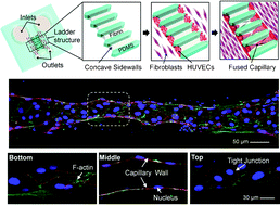
This paper describes the in vitro formation and characterization of perfusable capillary networks made of human umbilical vein endothelial cells (HUVECs) in microfluidic devices (MFDs). Using this platform, an array of three-dimensional (3D) tubular capillaries of various dimensions (50–150 μm in diameter and 100–1600 μm in length) can be formed reproducibly. To generate connected blood vessels, MFDs were completely filled with fibrin gel and subsequently processed to selectively leave behind gel structures inside the bridge channels. Following gel solidification, HUVECs were coated along the gel walls, on opposite ends of the patterned 3D fibrin gel. After 3–4 days, HUVECs migrating into the fibrin gel from opposite ends fused with each other, spontaneously forming a connected vessel that expressed tight junction proteins (e.g., ZO-1), which are characteristic of post-capillary venules. With ready access to a perfusable capillary network, we demonstrated perfusion of the vessels and imaged red blood cells (RBCs) and beads flowing through them. The results were reproducible (∼50% successful perfusable capillaries), consistent, and could be performed in a parallel manner (9 devices per well plate). Additionally, compatibility with high resolution live-cell microscopy and the possibility of incorporating other cell types makes this a unique experimental platform for investigating basic and applied aspects of angiogenesis, anastomosis, and vascular biology.

Graphical abstract: In vitro formation and characterization of a perfusable three-dimensional tubular capillary network in microfluidic devices

Supplementary files

Article information

Article type
Paper
Submitted
07 Feb 2012
Accepted
22 May 2012
First published
24 May 2012

Lab Chip, 2012,12, 2815-2822

In vitro formation and characterization of a perfusable three-dimensional tubular capillary network in microfluidic devices

J. H. Yeon, H. R. Ryu, M. Chung, Q. P. Hu and N. L. Jeon, Lab Chip, 2012, 12, 2815 DOI: 10.1039/C2LC40131B

To request permission to reproduce material from this article, please go to the Copyright Clearance Center request page.

If you are an author contributing to an RSC publication, you do not need to request permission provided correct acknowledgement is given.

If you are the author of this article, you do not need to request permission to reproduce figures and diagrams provided correct acknowledgement is given. If you want to reproduce the whole article in a third-party publication (excluding your thesis/dissertation for which permission is not required) please go to the Copyright Clearance Center request page.

Read more about how to correctly acknowledge RSC content.

Social activity

Spotlight

Advertisements