Issue 9, 2008

Millisecond denaturation dynamics of fluorescent proteins revealed by femtoliter container on micro-thermodevice

Abstract

Real-time observation of biomolecular behavior focusing on high speed temperature response is an essential endeavor for further biological study at the molecular level. This is because most of the important biological functions at the molecular level happen at the sub-second time scale. We used our own on-chip microheaters and microcontainers to observe the denaturation dynamics of fluorescent proteins at the millisecond time scale. The microheater controls the temperature in 1 ms under the microscope. Fluorescent proteins were contained in 28 fL PDMS microcontainers to prevent them from diffusing into the solution. The proteins were denatured by high temperatures and observed by a high speed CCD camera with 5 ms per frame. Hence, denaturation speeds of red fluorescent proteins (rDsRed and rHcRed) were measured to be 5–10 ms. Green fluorescent proteins (rAcGFP and rGFPuv) denatured with bi-exponential decay. rAcGFP denatured with time constants of 5 ms and 75 ms while rGFPuv denatured with 10 ms and 130 ms. This may be the reverse process of a two step renaturation of GFP observed in a previous report. This micro-thermodevice is applicable to other biomaterials such as nucleic acids or other proteins. It does not require any chemical treatment nor mutation to the biomaterial itself. Therefore, the methodology using this general purpose device gives access to biomolecular studies in short time scales and acts as a powerful tool in molecular biology.
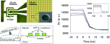
Graphical abstract: Millisecond denaturation dynamics of fluorescent proteins revealed by femtoliter container on micro-thermodevice

Article information

Article type
Communication
Submitted
21 Apr 2008
Accepted
15 Jul 2008
First published
29 Jul 2008
This article is Open Access

Lab Chip, 2008,8, 1436-1440

Millisecond denaturation dynamics of fluorescent proteins revealed by femtoliter container on micro-thermodevice

H. F. Arata, F. Gillot, T. Nojima, T. Fujii and H. Fujita, Lab Chip, 2008, 8, 1436 DOI: 10.1039/B806749J

This is an Open Access article. The full version of this article can be posted on a website/blog, posted on an intranet, photocopied, emailed, distributed in a course pack or distributed in Continuing Medical Education (CME) materials provided that it is not used for commercial purposes.

To request permission to reproduce material from this article in a commercial publication, please go to the Copyright Clearance Center request page.

If you are an author contributing to an RSC publication, you do not need to request permission provided correct acknowledgement is given.

If you are the author of this article, you do not need to request permission to reproduce figures and diagrams provided correct acknowledgement is given. If you want to reproduce the whole article in a third-party commercial publication (excluding your thesis/dissertation for which permission is not required) please go to the Copyright Clearance Center request page.

Read more about how to correctly acknowledge RSC content.

Social activity

Spotlight

Advertisements