
Optimisation of a laser ablation cell for detection of hetero-elements in proteins blotted onto membranes by use of inductively coupled plasma mass spectrometry
Abstract
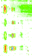
For imaging of elemental distribution in protein spots blotted onto membranes with a conventional size, we have investigated three different laser ablation cell geometries to find a design of a cell showing a constantly high signal intensity across the cell and possessing short wash-out times. For this purpose the resulting intensity after optimisation (He carrier gas flow rate, argon make-up gas, generator forward power) was measured for single laser shots at different positions on a steel metal sheet and for continuous ablation of nitrocellulose (NC) membranes during linear translation of the cell. In the latter experiment, ink bar structures have been printed on the NC membranes to simulate blotted protein spots and used to evaluate the local resolution and to determine an adequate translation velocity. The final cell has a volume of about 11.3 cm3 in which single shot signals are washed out in less than a second so that translation velocities of up to 1.5 mm s−1 can be applied to baseline separate structures with a distance of less than 2 mm, which will be demonstrated using an example of a phosphoprotein mixture separated by SDS-PAGE and blotted onto a NC membrane.
 Please wait while we load your content...

