Development of an ex vivo breast cancer lung colonization model utilizing a decellularized lung matrix†
Abstract
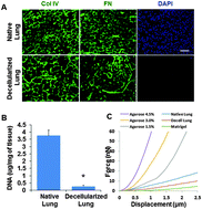
The metastatic spread of cancer cells to distant sites represents the major cause of cancer-related deaths in breast cancer patients, and lungs are one of the most common sites for metastatic colonization. Developing a physiologically relevant tissue culture model to mimic lung colonization of breast cancer is crucial for the investigation of the biology of cancer metastasis and evaluation of drug treatment efficacy. Here, we describe an ex vivo lung colonization assay for breast cancer using the native three-dimensional (3D) lung extracellular matrix. The native matrix was isolated from murine lungs using a decellularization technique, and the preservation of extracellular matrix (ECM) composition, integrity and mechanical properties was confirmed. We showed that metastatic MDA-MB 231 and 4T1 cells invaded and colonized in the decellularized lung matrix, whereas only a small mass of non-metastatic MCF7 cells survived under the same condition. Furthermore, knockdown of ZEB1, an epithelial–mesenchymal transition (EMT) inducer, significantly reduced invasion and colonization of MDA-MB 231 cells in the decellularized lung, suggesting an important role of EMT in breast cancer metastasis. We conclude that the decellularized lung retains the biophysical and biochemical properties of the lung ECM and provides a powerful tool to investigate the lung colonization of breast cancer.


 Please wait while we load your content...