An energy transfer kinetic probe for OH-quenchers in the Nd3+:YPO4 nanocrystals suitable for imaging in the biological tissue transparency window
Abstract
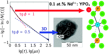
Tetragonal xenotime-type yttrium orthophosphate (YPO4) Nd3+ doped nanoparticles suitable for biomedical applications were prepared by microwave-hydrothermal treatment. We applied the energy transfer probing based on the analysis of kinetics of impurity quenching to determine the presence and spatial position of –OH fluorescence quenching acceptors in the impurity-containing nanoparticles. We show that the impurity quenching kinetics of the 0.1 at% Nd3+ doped YPO4 nanoparticles is a two stage (ordered and disordered) static kinetics, determined by a direct energy transfer to the –OH acceptors. Analyzing the ordered stage, we assume that the origin of the –OH groups is the protonation of the phosphate groups, while analyzing the disordered stage, we assume the presence of water molecules in the mesopores. We determine the dimension of the space of the –OH acceptors as d = 3 and quantify their absolute concentration using the disordered Förster stage of kinetics. We use the late stage of kinetics of fluorescence hopping (CDD ≫ CDA) quenching (the fluctuation asymptotics) at 1 at% Nd3+ concentration as an energy transfer probe to quantify the relative concentration of –OH molecular groups compared to an optically active rare-earth dopant in the volume of NPs, when energy migration over Nd3+ donors to the –OH acceptors accelerates fluorescence quenching. In doing so we use just one parameter  , defined by the relation of concentration of the –OH acceptors to the concentration of an optically active dopant. The higher is the α, the higher is the relative concentration of –OH acceptors in the volume of nanoparticles. We find α = 2.95 for the 1 at% Nd3+:YPO4 NPs that, according to the equation for α, and the results obtained for the values of the microparameters CDD(Nd–Nd) = 24.6 nm6 ms−1 and CDA(Nd–OH) = 0.6 nm6 ms−1, suggests twenty times higher concentration for acceptors other than donors. As the main result we have established that the majority of –OH acceptors is located not on the surface of the Nd3+:YPO4 nanoparticles, as many researchers assumed, but in their volume, and can be either associated with crystal structure defects or located in the mesopores.


 Please wait while we load your content...