Issue 6, 2013

Structural prediction of a rhodamine-based biosensor and comparison with biophysical data

Abstract

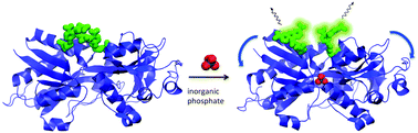
The predicted structure has been calculated for a protein-based biosensor for inorganic phosphate (Pi), previously developed by some of us (Okoh et al., Biochemistry, 2006, 45, 14764). This is the phosphate binding protein from Escherichia coli labelled with two rhodamine fluorophores. Classical molecular dynamics and hybrid Car–Parrinello/molecular mechanics simulations allow us to provide molecular models of the biosensor both in the presence and in the absence of Pi. In the latter case, the rhodamine fluorophores maintain a stacked conformation in a ‘face A to face B’ orientation, which is different from the ‘face A to face A’ stacked orientation of free fluorophores in aqueous solution (Ilich et al., Spectrochim. Acta, Part A, 1996, 52, 1323). A protein conformation change upon binding Pi prevents significant stacking of the two rhodamines. In both states, the rhodamine fluorophores form hydrophobic contact with LEU291, without establishing significant hydrogen bonds with the protein. The accuracy of the models is established by a comparison between calculated and experimental absorption and circular dichroism spectra.

Graphical abstract: Structural prediction of a rhodamine-based biosensor and comparison with biophysical data

Supplementary files

Article information

Article type
Paper
Submitted
14 Jul 2012
Accepted
30 Nov 2012
First published
17 Dec 2012

Phys. Chem. Chem. Phys., 2013,15, 2177-2183

Structural prediction of a rhodamine-based biosensor and comparison with biophysical data

M. B. Gonçalves, J. Dreyer, P. Lupieri, C. Barrera-Patiño, E. Ippoliti, M. R. Webb, J. E. T. Corrie and P. Carloni, Phys. Chem. Chem. Phys., 2013, 15, 2177 DOI: 10.1039/C2CP42396K

To request permission to reproduce material from this article, please go to the Copyright Clearance Center request page.

If you are an author contributing to an RSC publication, you do not need to request permission provided correct acknowledgement is given.

If you are the author of this article, you do not need to request permission to reproduce figures and diagrams provided correct acknowledgement is given. If you want to reproduce the whole article in a third-party publication (excluding your thesis/dissertation for which permission is not required) please go to the Copyright Clearance Center request page.

Read more about how to correctly acknowledge RSC content.

Social activity

Spotlight

Advertisements