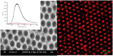
Gold nanocavity arrays were prepared on fluorine-doped tin oxide on glass by electrochemical deposition of gold through monolayers of polystyrene spheres. The impact of the resulting spherical cap architecture on the photophysics of solutions and self-assembled monolayers of luminophore encapsulated within the nanocavities is reported for the first time. From conventional confocal fluorescence microscopy, the emission intensity of solutions of [Ru(bpy)2(Qbpy)]2+ (where bpy is 2,2′-bipyridyl and Qbpy is 2,2′:4,4″:4,4″-quarterpyridyl) and fullerene (C60) encapsulated within the 820 nm diameter nanocavities was demonstrated to increase by approximately an order of magnitude compared with that of the associated bulk solution. Comparison was also made with the emission observed for luminophore solution encapsulated in cobalt nanocavities of comparable dimensions, where plasmonic interactions are not anticipated. Again, approximately an order of magnitude enhancement was observed for the gold arrays. Luminescence lifetime imaging revealed that the enhancement of the emission intensity of this solution within the nanocavity was accompanied by a small but significant decrease in the luminescent lifetime for [Ru(bpy)2(Qbpy)]2+. Enhancement was, in addition, strongly influenced by the wavelength of excitation, suggesting that plasmonics may play a role in the enhancement of the excitation process. An important observation from confocal imaging studies was that the dimensions of the luminophore emission field from solution within the cavities were significantly smaller than the dimensions of the cavity aperture and corresponded to a little more than that of the point spread function of the microscope. This indicates that its origin is significantly smaller than the wavelength of the emitted light and suggests that luminescence enhancement is highly localised. When the array was filled with a solution of [Ru(bpy)2(Qbpy)]2+ the emission spectrum of this complex was red shifted and broadened compared with that of the bulk solution, typical of the formation of a luminescent surface film. In addition, significant enhancement was only observed when the solution was sonicated into the array. Both these observations suggest that the emission enhancement is localised near the bottom of the cavity. Self-assembled monolayers of [Ru(bpy)2(Qbpy)]2+ were formed on the array and approximately 7 orders of magnitude enhancement of the Raman signal was achieved. Significantly, the emission intensity was approximately 4-fold higher for the monolayer than for a solid film under the same conditions, but surface quenching is thought to play a significant role in the observed decrease in lifetime for the monolayer of this complex on the array.
You have access to this article
 Please wait while we load your content...
Something went wrong. Try again?
Please wait while we load your content...
Something went wrong. Try again?


