Issue 18, 2014

X-ray chemical imaging and the electronic structure of a single nanoplatelet Ni/graphene composite

Abstract

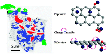
Chemical imaging and quantitative analysis of a single graphene nanoplatelet grown with Ni nanoparticles (Ni/graphene) has been performed by scanning transmission X-ray microscopy (STXM). Local electronic and chemical structure of Ni/graphene has been investigated by spatially resolved C, O K-edges and Ni L-edge X-ray absorption near edge structure (XANES) spectroscopy, revealing the covalent anchoring of Ni(0) on graphene. This study facilitates the understanding of the structure modification of host materials for hydrogen storage and offers a better understanding of interaction between Ni particles and graphene.

Graphical abstract: X-ray chemical imaging and the electronic structure of a single nanoplatelet Ni/graphene composite

Supplementary files

Article information

Article type
Communication
Submitted
13 Sep 2013
Accepted
02 Jan 2014
First published
06 Jan 2014

Chem. Commun., 2014,50, 2282-2285

Author version available

X-ray chemical imaging and the electronic structure of a single nanoplatelet Ni/graphene composite

C. Zhou, J. Wang and J. A. Szpunar, Chem. Commun., 2014, 50, 2282 DOI: 10.1039/C3CC47008C

To request permission to reproduce material from this article, please go to the Copyright Clearance Center request page.

If you are an author contributing to an RSC publication, you do not need to request permission provided correct acknowledgement is given.

If you are the author of this article, you do not need to request permission to reproduce figures and diagrams provided correct acknowledgement is given. If you want to reproduce the whole article in a third-party publication (excluding your thesis/dissertation for which permission is not required) please go to the Copyright Clearance Center request page.

Read more about how to correctly acknowledge RSC content.

Social activity

Spotlight

Advertisements