Issue 4, 2002

Three-dimensional imaging of YB56 by high-resolution electron microscopy

Abstract

The three-dimensional potential map of YB56 was obtained by inverse Fourier transformation of three-dimensional phases and amplitudes in three high-resolution images taken along the [100], [110] and [111] directions of YB56 crystals; the size of the imaging region was 14 nm × 14 nm × ∼4 nm, and the image directly showed the three-dimensional potential map of the crystal, a useful method for three-dimensional structure analysis in nanoscale regions.
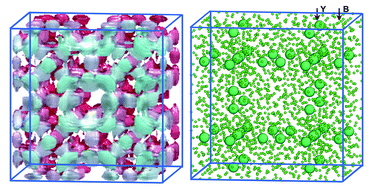
Graphical abstract: Three-dimensional imaging of YB56 by high-resolution electron microscopy

Supplementary files

Article information

Article type
Communication
Submitted
05 Sep 2001
Accepted
06 Dec 2001
First published
18 Jan 2002

Chem. Commun., 2002, 302-303

Three-dimensional imaging of YB56 by high-resolution electron microscopy

T. Oku, Chem. Commun., 2002, 302 DOI: 10.1039/B107864J

To request permission to reproduce material from this article, please go to the Copyright Clearance Center request page.

If you are an author contributing to an RSC publication, you do not need to request permission provided correct acknowledgement is given.

If you are the author of this article, you do not need to request permission to reproduce figures and diagrams provided correct acknowledgement is given. If you want to reproduce the whole article in a third-party publication (excluding your thesis/dissertation for which permission is not required) please go to the Copyright Clearance Center request page.

Read more about how to correctly acknowledge RSC content.

Social activity

Spotlight

Advertisements