The surrounding tissue contributes to smooth muscle cells’ regeneration and vascularization of small diameter vascular grafts†
 *a
and
Deling
Kong
*ab
*a
and
Deling
Kong
*ab
Abstract
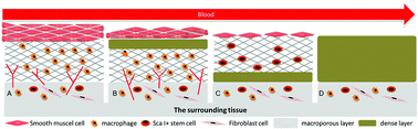
Small diameter vascular grafts have been promising substitutes for bypass surgery to treat cardiovascular disease. However, no ideal product is available in the clinic. In order to design improved, next generation vascular grafts, it is essential to understand the cellular and molecular mechanisms underlying tissue regeneration after vascular graft implantation. Two diverse microenvironments, circulating blood and the surrounding tissue, are involved in the regeneration process after vascular graft implantation in situ. However, their regenerative functions are not completely understood. To elucidate their roles in regeneration, we used electrospinning to fabricate four types of tubular scaffolds with a structure consisting of a microfiber layer (fiber diameter ∼ 6 μm) and a nanofiber layer (fiber diameter < 1 μm): microfiber scaffold, nanofiber scaffold, outer microfiber bilayer scaffold and inner microfiber bilayer scaffold. In the outer microfiber scaffold, cells from the surrounding tissue were allowed into the scaffold but not cells from the circulating blood while it was opposite in the inner microfiber scaffold. The processes of endothelium formation, smooth muscle cell regeneration, neo-tissue formation and vascularization of these scaffolds were analyzed with a rat left common carotid artery replacement model. Our data showed that smooth muscle cells’ regeneration and vascularization were different among the four types of scaffolds. The thickest neo-tissue and α-SMA+ cell layers were detected in the microfiber scaffold group while the thinnest in the nanofiber scaffold group, and thicker neo-tissue and α-SMA+ cell layers were found in the outer microfiber bilayer scaffold group compared to the inner microfiber bilayer scaffold group. In addition, vascularization in the outer microfiber bilayer scaffold group and microfiber group was dramatically better than the inner microfiber bilayer scaffold group and the nanofiber group. Furthermore, we demonstrated that the regenerated SMCs were associated with the CD206+ macrophages in the graft wall. In all, the microfiber scaffold showed the best neo-tissue regeneration in vivo. These results indicate that the surrounding tissue contributes more to vascular regeneration than circulating blood. This finding gives a significant design clue that modulating the vascular surrounding tissue will be an alternative strategy for designing advanced and feasible small diameter vascular grafts.
 Please wait while we load your content...

