A de-waxing methodology for scanning probe microscopy
 a
Peter
Gardner,
c
Steve D.
Barrett,
a
Asterios
Triantafyllou,
d
Janet M.
Risk
b
and
Peter
Weightman
*a
a
Peter
Gardner,
c
Steve D.
Barrett,
a
Asterios
Triantafyllou,
d
Janet M.
Risk
b
and
Peter
Weightman
*a
Abstract
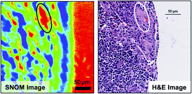
A de-waxing protocol that successfully removes paraffin from tissue microarray (TMA) cores of fixed tissue obtained from oral cancer is described. The success of the protocol is demonstrated by the comparison of Fourier transform infrared (FTIR) results obtained on paraffin-embedded and de-waxed tissue and the absence of any significant correlations between infrared scanning near-field optical microscopy (SNOM) images of de-waxed tissue obtained at the three main paraffin IR peaks. The success of the protocol in removing paraffin from tissue is also demonstrated by images obtained with scanning electron microscopy (SEM) and by energy dispersive spectra (EDS) of a de-waxed CaF2 disc which shows no significant contribution from carbon. The FTIR spectra of the de-waxed TMA core overlaps that obtained from OE19 oesophageal cancer cells which had never been exposed to paraffin.
 Please wait while we load your content...

