Quantitation of acute monocytic leukemia cells spiked in control monocytes using surface-enhanced Raman spectroscopy
Abstract
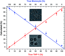
Surface enhanced Raman spectroscopy (SERS) was used to quantify leukemia cells spiked in control cells. The novelty of the technique lies in preparing cell lysates by ultrasound sonication and mixing with silver nanoparticles which allow reproducible interaction of biomolecules and nanoparticles. The SERS spectra of these mixtures not only exhibit enhanced bands of intracellular proteins and nucleic acids, but also spectral variations for accurate cell identification. Here, samples from an acute monocytic leukemia cell line, THP-1, and control monocytes from three donors served as the in vitro model system for leukemia. For quantitative analysis, seven mixtures containing different percentile amounts of leukemia lysates and lysates from control monocytes were prepared ranging from 0% to 100% and SERS spectra were measured. The more intense spectral contributions of proteins relative to nucleic acids correlated with the larger cytoplasm to nucleus ratio of leukemia cells than control monocytes. The experimental SERS spectra were fitted by a non-negative least squares (NNLS) algorithm to calculate the percentile amounts of each of the cell types and to determine their contributions to the mixtures. Even in a mixture with control monocytes (360 μl), a small amount (5 μl) of leukemia cells was detected, which represents 10% of leukemia cells considering their twofold larger diameter and eightfold larger volume. As this value is well below the threshold of 20% blast cells for leukemia diagnosis, this approach is very promising for both qualitative and quantitative analysis of human cell mixtures. This study demonstrates the potential of SERS and NNLS fitting as a rapid method for diagnosis of acute monocytic leukemia in human blood or bone marrow samples because only a single spectrum is required.


 Please wait while we load your content...