Issue 2, 2020

A simple microfluidic device for live-imaging of the vertical section of epithelial cells

Abstract

We investigated the capability of simple microfluidic devices with trenches having vertical sidewalls for live-cell fluorescence imaging of adherent cells. An epithelial cell line that forms a two-dimensional (2D) sheet was cultured to adhere to the vertical sidewall so that its vertical section can be imaged directly using ordinal inverted-type laser-scanning microscopy. The material and the structure of the device were characterized. We show that the detailed distribution of intracellular organelles, such as microtubules and mitochondria, and of intercellular apparatus, such as claudin and zonula occludens, can be imaged with high spatio-temporal resolution with a single scan.
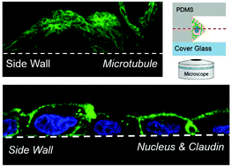

Graphical abstract: A simple microfluidic device for live-imaging of the vertical section of epithelial cells

Supplementary files

Article information

Article type
Paper
Submitted
28 Oct 2019
Accepted
09 Nov 2019
First published
11 Nov 2019

Analyst, 2020,145, 667-674

A simple microfluidic device for live-imaging of the vertical section of epithelial cells

S. Araki, M. Nakano, M. Tsugane, F. Sunaga, M. Hattori, M. Nakano, T. Nagai and H. Suzuki, Analyst, 2020, 145, 667 DOI: 10.1039/C9AN02165E

To request permission to reproduce material from this article, please go to the Copyright Clearance Center request page.

If you are an author contributing to an RSC publication, you do not need to request permission provided correct acknowledgement is given.

If you are the author of this article, you do not need to request permission to reproduce figures and diagrams provided correct acknowledgement is given. If you want to reproduce the whole article in a third-party publication (excluding your thesis/dissertation for which permission is not required) please go to the Copyright Clearance Center request page.

Read more about how to correctly acknowledge RSC content.

Social activity

Spotlight

Advertisements