
Accurate assessment of liver steatosis in animal models using a high throughput Raman fiber optic probe
Abstract
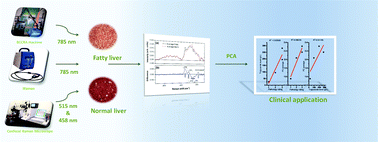
Due to the shortage of healthy donor organs, steatotic livers are commonly used for transplantation, placing patients at higher risk for graft dysfunction and lower survival rates. Raman Spectroscopy is a technique which has shown the ability to rapidly detect the vibration state of C–H bonds in triglycerides. The aim of this study is to determine whether conventional Raman spectroscopy can reliably detect and quantify fat in an animal model of liver steatosis. Mice and rats fed a methionine and choline-deficient (MCD) and control diets were sacrificed on one, two, three and four weeks’ time points. A confocal Raman microscope, a commercial Raman (iRaman) fiber optic probe and a highly sensitive Raman fiber optic probe system, the latter utilizing a 785 nm excitation laser, were used to detect changes in the Raman spectra of steatotic mouse livers. Thin layer chromatography was used to assess the triglyceride content of liver specimens, and sections were scored blindly for fat content using histological examination. Principal component analysis (PCA) of Raman spectra was used to extract the principal components responsible for spectroscopic differences with MCD week (time on MCD diet). Confocal Raman microscopy revealed the presence of saturated fats in mice liver sections. A commercially available handheld Raman spectroscopy probe could not distinguish the presence of fat in the liver whereas our specially designed, high throughput Raman system could clearly distinguish lobe-specific changes in fat content. In the left lobe in particular, the Raman PC scores exhibited a significant correlation (R2 = 0.96) with the gold standard, blinded scoring by histological examination. The specially designed, high throughput Raman system can be used for clinical purposes. Its application to the field of transplantation would enable surgeons to determine the hepatic fat content of the donor's liver in the field prior to proceeding with organ retrieval. Next steps include validating these results in a prospective analysis of human liver transplantation implant biopsies.
 Please wait while we load your content...

