Issue 9, 2009

Cu2+-selective naked-eye and fluorescent probe: its crystal structure and application in bioimaging

Abstract

A new fluorescent and colorimetric Cu2+ probe was synthesized, and realized optical imaging in RAW264.7 macrophages. The design strategy was based on a change in structure between spirocyclic (non-fluorescent) and ring-open (fluorescent) forms of rhodamine-based dyes, and its crystal structure was presented to explain the binding mode. Upon treatment with Cu2+, the weakly fluorescent probe exhibited a strong fluorescence response, high selectivity and was quantitative for Cu2+ under physiological conditions. In addition, the off–on-type fluorescent change upon the addition of Cu2+ was also applied in bioimaging.
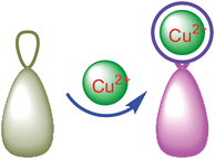
Graphical abstract: Cu2+-selective naked-eye and fluorescent probe: its crystal structure and application in bioimaging

Supplementary files

Article information

Article type
Paper
Submitted
14 Jan 2009
Accepted
02 Jul 2009
First published
17 Jul 2009

Analyst, 2009,134, 1826-1833

Cu2+-selective naked-eye and fluorescent probe: its crystal structure and application in bioimaging

F. Yu, W. Zhang, P. Li, Y. Xing, L. Tong, J. Ma and B. Tang, Analyst, 2009, 134, 1826 DOI: 10.1039/B823360H

To request permission to reproduce material from this article, please go to the Copyright Clearance Center request page.

If you are an author contributing to an RSC publication, you do not need to request permission provided correct acknowledgement is given.

If you are the author of this article, you do not need to request permission to reproduce figures and diagrams provided correct acknowledgement is given. If you want to reproduce the whole article in a third-party publication (excluding your thesis/dissertation for which permission is not required) please go to the Copyright Clearance Center request page.

Read more about how to correctly acknowledge RSC content.

Social activity

Spotlight

Advertisements