
Fluorescently-tagged magnetic protein nanoparticles for high-resolution optical and ultra-high field magnetic resonance dual-modal cerebral angiography†

Abstract
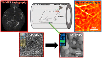
Extremely small paramagnetic iron oxide nanoparticles (FeMNPs) (<5 nm) can enhance positive magnetic resonance imaging (MRI) contrast by shortening the longitudinal relaxation time of water (T1), but these nanoparticles experience rapid renal clearance. Here, magnetic protein nanoparticles (MPNPs) are synthesized from protein-conjugated citric acid coated FeMNPs (c-FeMNPs) without loss of the T1 MRI properties and tagged with fluorescent dye (f-MPNPs) for optical cerebrovascular imaging. The c-FeMNPs shows average size 3.8 ± 0.7 nm with T1 relaxivity (r1) of 1.86 mM−1 s−1 and transverse/longitudinal relaxivity ratio (r2/r1) of 2.53 at 11.7 T. The f-MPNPs show a higher r1 value of 2.18 mM−1 s−1 and r2/r1 ratio of 2.88 at 11.7 T, which generates excellent positive MRI contrast. In vivo cerebral angiography with f-MPNPs enables detailed microvascular contrast enhancement for differentiation of major blood vessels of murine brain, which corresponds well with whole brain three-dimensional time-of-flight MRI angiograms (17 min imaging time with 60 ms repetition time and 40 μm isotropic voxels). The real-time fluorescence angiography enables unambiguous detection of brain capillaries with diameter < 40 μm. Biodistribution examination revealed that f-MPNPs were safely cleared by the organs like the liver, spleen, and kidneys within a day after injection. Blood biochemical assays demonstrated no risk of iron overload in both rats and mice. With hybrid neuroimaging technologies (e.g., MRI-optical) on the rise, f-MPNPs built on this platform can generate exciting neuroscience applications.
 Please wait while we load your content...

