Live-cell imaging of the nucleolus and mapping mitochondrial viscosity with a dual function fluorescent probe†
Tarushyam
Mukherjee,  a
Virupakshi
Soppina,
*b
Yves
Mély,
c
Andrey S.
Klymchenko,
c
Mayeul
Collot
c
and
Sriram
Kanvah
*a
a
Virupakshi
Soppina,
*b
Yves
Mély,
c
Andrey S.
Klymchenko,
c
Mayeul
Collot
c
and
Sriram
Kanvah
*a
a
Virupakshi
Soppina,
*b
Yves
Mély,
c
Andrey S.
Klymchenko,
c
Mayeul
Collot
c
and
Sriram
Kanvah
*a
Abstract
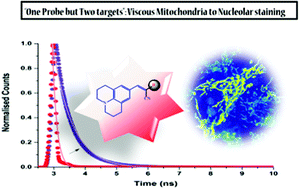
Visualization of sub-cellular organelles allows the determination of various cellular processes and the underlying mechanisms. Herein, we report a fluorescent probe, bearing push–pull substituents emitting at 600 nm and its application in cellular imaging. The probe shows dual imaging of mitochondria and nucleoli and maps mitochondrial viscosity in live cells under various physiological variations and show minimum cytotoxicity. Nucleolar staining is confirmed by RNAase digestion.
- This article is part of the themed collection: Chemical Biology in OBC
 Please wait while we load your content...

