
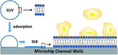
Supported bilayer membranes for reducing cell adhesion in microfluidic devices

Abstract
The high surface area-to-volume ratio of microfluidic channels makes them susceptible to fouling and clogging when used for biological analyses, including cell-based assays. We evaluated the role of electrostatic and van der Waals interactions in cell adhesion in PDMS microchannels coated with supported lipid bilayers and identified conditions that resulted in minimal cell adhesion. For low ionic strength buffer, optimum results were obtained for a zwitterionic coating of pure egg phosphatidylcholine; for a rich growth medium, the best results were obtained for zwitterionic bilayers or those with slight negative or moderate positive charge from the incorporation of 5–10 mol% egg phosphatidylglycerol or 30 mol% ethylphosphocholine. In both solutions, the presence of 10 g L−1 glucose in the cell suspension reduced cell adhesion. Under optimum conditions, all cells were consistently removed from the channels, demonstrating the utility of these coatings for whole-cell microfluidic assays. These results provide practical information for immediate application and suggest future research areas on cell–lipid interactions.
 Please wait while we load your content...

