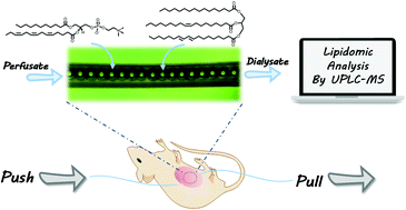
Open-flow microperfusion combined with mass spectrometry for in vivo liver lipidomic analysis†
Abstract
At present, conventional microdialysis (MD) techniques cannot efficiently sample lipids in vivo, possibly due to the high mass transfer resistance and/or the serious adsorption of lipids onto the semi-permeable membrane of a MD probe. The in vivo monitoring of lipids could be of great significance for the study of disease development and mechanisms. In this work, an open-flow microperfusion (OFM) probe was fabricated, and the conditions for sampling lipids via OFM were optimized. Using OFM, the recovery of lipid standards was improved to more than 34.7%. OFM is used for the in vivo sampling of lipids in mouse liver tissue with fibrosis, and it is then combined with mass spectrometry (MS) to perform lipidomic analysis. 156 kinds of lipids were identified in the dialysate collected via OFM, and it was found that the phospholipid levels, including PC, PE, and SM, were significantly higher in a liver suffering from fibrosis. For the first time, OFM combined with MS to sample and analyze lipids has provided a promising platform for in vivo lipidomic studies.


 Please wait while we load your content...