Issue 55, 2020

Binding modes of thioflavin T and Congo red to the fibril structure of amyloid-β(1–42)

Abstract

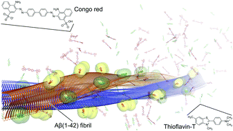
Binding modes for the amyloid-β(1–42) fibril fluorescent dyes thioflavin T and Congo red were predicted by molecular dynamics simulations and binding free energy calculations. Both probes bind on the fibril surface to primarily hydrophobic grooves, with their long axis oriented almost parallel to the fibril axis. The computed binding affinities are in agreement with experimental values. The binding modes also explain observables from previous structural studies and, thus, provide a starting point for the systematic search and design of novel molecules, which may improve in vitro diagnostics for Alzheimer's disease.

Graphical abstract: Binding modes of thioflavin T and Congo red to the fibril structure of amyloid-β(1–42)

Supplementary files

Article information

Article type
Communication
Submitted
17 Feb 2020
Accepted
01 Jun 2020
First published
01 Jun 2020

Chem. Commun., 2020,56, 7589-7592

Binding modes of thioflavin T and Congo red to the fibril structure of amyloid-β(1–42)

B. Frieg, L. Gremer, H. Heise, D. Willbold and H. Gohlke, Chem. Commun., 2020, 56, 7589 DOI: 10.1039/D0CC01161D

To request permission to reproduce material from this article, please go to the Copyright Clearance Center request page.

If you are an author contributing to an RSC publication, you do not need to request permission provided correct acknowledgement is given.

If you are the author of this article, you do not need to request permission to reproduce figures and diagrams provided correct acknowledgement is given. If you want to reproduce the whole article in a third-party publication (excluding your thesis/dissertation for which permission is not required) please go to the Copyright Clearance Center request page.

Read more about how to correctly acknowledge RSC content.

Social activity

Spotlight

Advertisements