Osteogenic potential of Zn2+-passivated carbon dots for bone regeneration in vivo†
Abstract
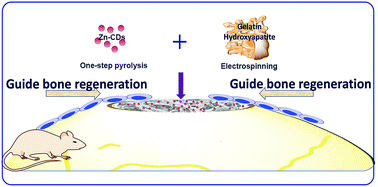
Carbon dots are a new kind of nanomaterial which has great potential in biomedical applications. Previously, we have synthesized novel Zn2+-passivated carbon dots (Zn-CDs) which showed good osteogenic activity in vitro. In this study, we will further investigate the osteogenic effects of Zn-CDs in vivo which is essential before their clinical use. Herein, Zn2+-passivated carbon dots (Zn-CDs) are prepared and characterized as previously reported. Then, the optimum dose for inducing osteoblasts was evaluated by MTS assay, intracellular reactive oxygen species (ROS) detection, alkaline phosphatase (ALP) activity test and alizarin red staining in vitro. Finally, a 5 mm diameter calvarial bone defect model was created in rats and Zn-CDs were applied for repairing the critical bone defect. It was shown that zinc gluconate (Zn-G) and Zn-CDs promoted the survival of bone marrow stromal cells (BMSCs) when the zinc ion concentration was 10−4 mol L−1 (Zn-G: 45.6 μg mL−1) and 10−5 mol L−1 (Zn-CDs: 300 μg mL−1) or below respectively. With regard to the osteogenic capability, the ALP activity induced by Zn-CDs was significantly higher than that by Zn-G. Besides, the results of alizarin red staining showed that the area of calcified nodules was increased in a dose-dependent manner in the Zn-CD group. Moreover, there were more calcium nodules in the Zn-CD group than in the Zn-G group at the same concentration of Zn2+ (10−5 mol L−1). Taken together, Zn-CDs achieved the highest osteogenic effect at the concentration of 10−5 mol L−1 without affecting cell proliferation in long-term stimulation. Importantly, the volume of new bone formation in the Zn-CD group (6.66 ± 1.25 mm3) was twice higher than that in the control group (3.33 ± 0.94 mm3) in vivo. Further histological evaluation confirmed the markedly new bone formation at 8 weeks in the Zn-CD group. The in vitro and in vivo experiments revealed that Zn-CDs could be a new predictable nanomaterial with good biocompatibility and fluorescence properties for guiding bone regeneration.


 Please wait while we load your content...