Issue 15, 2019

An in cellulo-activated multicolor cell labeling approach used to image dying cell clearance

Abstract

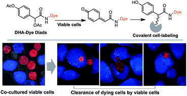
Dying cell clearance is critical for myriad biological processes such as tissue homeostasis. We herein report an enzyme-activated fluorescence cell labeling approach and its use for multicolor imaging of dying cell clearance. Diacetylated 4-hydroxymandelic acid (DHA)-conjugated dyes give rise to reactive quinone methides upon deacetylation in live cells, which in turn covalently labels cellular proteins. With partner cells tagged with distinct fluorescence, apoptotic cell clearance by Raw 264.7 macrophages and epithelial HeLa cells was captured by confocal microscopy, showing the potential of DHA-based cell labeling for investigating cell–cell interactions.

Graphical abstract: An in cellulo-activated multicolor cell labeling approach used to image dying cell clearance

Supplementary files

Article information

Article type
Paper
Submitted
17 May 2019
Accepted
11 Jun 2019
First published
18 Jun 2019

Analyst, 2019,144, 4687-4693

An in cellulo-activated multicolor cell labeling approach used to image dying cell clearance

Y. Shi, R. Zhu, Z. Xue, J. Han and S. Han, Analyst, 2019, 144, 4687 DOI: 10.1039/C9AN00904C

To request permission to reproduce material from this article, please go to the Copyright Clearance Center request page.

If you are an author contributing to an RSC publication, you do not need to request permission provided correct acknowledgement is given.

If you are the author of this article, you do not need to request permission to reproduce figures and diagrams provided correct acknowledgement is given. If you want to reproduce the whole article in a third-party publication (excluding your thesis/dissertation for which permission is not required) please go to the Copyright Clearance Center request page.

Read more about how to correctly acknowledge RSC content.

Social activity

Spotlight

Advertisements