
Through tissue imaging of a live breast cancer tumour model using handheld surface enhanced spatially offset resonance Raman spectroscopy (SESORRS)†
 a
Lauren E.
Jamieson,
a
Samuel
Mabbott,
a
Konstantinos
Plakas,
b
Neil C.
Shand,
c
Michael R.
Detty,
b
Duncan
Graham
a
and
Karen
Faulds
*a
a
Lauren E.
Jamieson,
a
Samuel
Mabbott,
a
Konstantinos
Plakas,
b
Neil C.
Shand,
c
Michael R.
Detty,
b
Duncan
Graham
a
and
Karen
Faulds
*a
Abstract
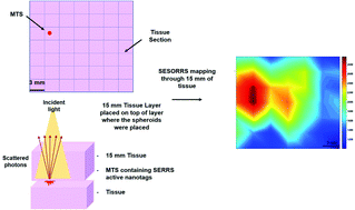
In order to improve patient survival and reduce the amount of unnecessary and traumatic biopsies, non-invasive detection of cancerous tumours is of imperative and urgent need. Multicellular tumour spheroids (MTS) can be used as an ex vivo cancer tumour model, to model in vivo nanoparticle (NP) uptake by the enhanced permeability and retention (EPR) effect. Surface enhanced spatially offset Raman spectroscopy (SESORS) combines both surface enhanced Raman spectroscopy (SERS) and spatially offset Raman spectroscopy (SORS) to yield enhanced Raman signals at much greater sub-surface levels. By utilizing a reporter that has an electronic transition in resonance with the laser frequency, surface enhanced resonance Raman scattering (SERRS) yields even greater enhancement in Raman signal. Using a handheld SORS spectrometer with back scattering optics, we demonstrate the detection of live breast cancer 3D MTS containing SERRS active NPs through 15 mm of porcine tissue. False color 2D heat intensity maps were used to determine tumour model location. In addition, we demonstrate the tracking of SERRS-active NPs through porcine tissue to depths of up to 25 mm. This unprecedented performance is due to the use of red-shifted chalcogenpyrylium-based Raman reporters to demonstrate the novel technique of surface enhanced spatially offset resonance Raman spectroscopy (SESORRS) for the first time. Our results demonstrate a significant step forward in the ability to detect vibrational fingerprints from a tumour model at depth through tissue. Such an approach offers significant promise for the translation of NPs into clinical applications for non-invasive disease diagnostics based on this new chemical principle of measurement.
- This article is part of the themed collection: Advances in Optical and Electrochemical Techniques for Biomedical Imaging
 Please wait while we load your content...

