
Enhanced magnetic microcytometer with 3D flow focusing for cell enumeration†
 *ab
Marco
Martins,
a
Lester C.
Barnsley,
cd
Bruno F. B.
Silva,
a
Susana
Cardoso,
e
Lorena
Diéguez,
a
Begoña
Espiña
a
and
Paulo P.
Freitas
abe
*ab
Marco
Martins,
a
Lester C.
Barnsley,
cd
Bruno F. B.
Silva,
a
Susana
Cardoso,
e
Lorena
Diéguez,
a
Begoña
Espiña
a
and
Paulo P.
Freitas
abe
Abstract
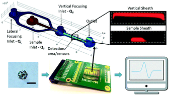
We report the design and characterization of a lateral and vertical hydrodynamic focusing feature for whole cell detection on a miniaturized flow cytometer. The developed system, based on magnetic sensing, incorporates spin valve sensors on the bottom of the microfluidic channels that detect cells labeled with magnetic beads. An adaptable 3D hydrodynamic focusing system was developed that pushes labeled cells towards the bottom of the microchannel, closer to the sensors, allowing increased signal amplitude for cells labeled with magnetic beads and enhanced discrimination of labeled cells. Fluorescence microscopy indicates that the lateral and vertical hydrodynamic focusing effect was adequately implemented, consistent with simulation predictions. The sensitivity of the system to detect labeled cells was improved by at least two-fold. By estimating the coverage of magnetic beads on cells, the signal from labeled cells could be predicted using a mathematical model, which also demonstrated the sensitivity of the signal to the height of the cells relative to the sensor. The system is versatile allowing interchangeable flow rates for cells with different diameters.
 Please wait while we load your content...

