Quantitative temporal interrogation in 3D of bioengineered human cartilage using multimodal label-free imaging†
Abstract
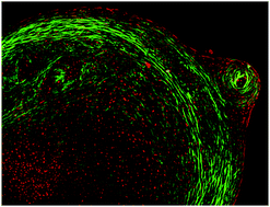
The unique properties of skeletal stem cells have attracted significant attention in the development of strategies for skeletal regeneration. However, there remains a crucial unmet need to develop quantitative tools to elucidate skeletal cell development and monitor the formation of regenerated tissues using non-destructive techniques in 3D. Label-free methods such as coherent anti-Stokes Raman scattering (CARS), second harmonic generation (SHG) and two-photon excited auto-fluorescence (TPEAF) microscopy are minimally invasive, non-destructive, and present new powerful alternatives to conventional imaging techniques. Here we report a combination of these techniques in a single multimodal system for the temporal assessment of cartilage formation by human skeletal cells. The evaluation of bioengineered cartilage, with a new parameter measuring the amount of collagen per cell, collagen fibre structure and chondrocyte distribution, was performed using the 3D non-destructive platform. Such 3D label-free temporal quantification paves the way for tracking skeletal cell development in real-time and offers a paradigm shift in tissue engineering and regenerative medicine applications.


 Please wait while we load your content...