
Potential use of MCR-ALS for the identification of coeliac-related biochemical changes in hyperspectral Raman maps from pediatric intestinal biopsies†

Abstract
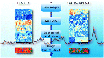
Raman hyperspectral imaging is an emerging practice in biological and biomedical research for label free analysis of tissues and cells. Using this method, both spatial distribution and spectral information of analyzed samples can be obtained. The current study reports the first Raman microspectroscopic characterisation of colon tissues from patients with Coeliac Disease (CD). The aim was to assess if Raman imaging coupled with hyperspectral multivariate image analysis is capable of detecting the alterations in the biochemical composition of intestinal tissues associated with CD. The analytical approach was based on a multi-step methodology: duodenal biopsies from healthy and coeliac patients were measured and processed with Multivariate Curve Resolution Alternating Least Squares (MCR-ALS). Based on the distribution maps and the pure spectra of the image constituents obtained from MCR-ALS, interesting biochemical differences between healthy and coeliac patients has been derived. Noticeably, a reduced distribution of complex lipids in the pericryptic space, and a different distribution and abundance of proteins rich in beta-sheet structures was found in CD patients. The output of the MCR-ALS analysis was then used as a starting point for two clustering algorithms (k-means clustering and hierarchical clustering methods). Both methods converged with similar results providing precise segmentation over multiple Raman images of studied tissues.
 Please wait while we load your content...