
Universally applicable three-dimensional hydrodynamic focusing in a single-layer channel for single cell analysis†

Abstract
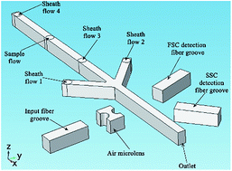
Three-dimensional (3D) hydrodynamic focusing of particles/cells is an important aspect of microfluidic cytometers to guarantee counting accuracy and measurement stability. In this article, a microfluidic flow cytometry system which integrated 3D hydrodynamic focusing and integrated on-chip optical systems on a single-layer microfluidic structure was demonstrated. By systematically adjusting the flow rate of the four-way sheath and sample flows, particles were hydrodynamically focused in horizontal and vertical directions to form a single-file flow in the middle of the microchannel. The 3D hydrodynamic focusing using only a single-layer microfluidic structure was verified by computational fluid dynamics numerical simulations and experiments. The 3D focusing experimental results of rhodamine solution indicated that the sample flow had been hydrodynamically focused in 3D. Furthermore, flow cytometric detections of polystyrene particles and human basophils were performed using the developed device. The measurement results demonstrated that the microfluidic cytometer with 3D hydrodynamic focusing exhibited comparable performance to a commercial cytometer. Consequently, the microfluidic cytometer developed in this article possesses the advantages of simple fabrication, precise 3D hydrodynamic focusing, and on-chip optical signal detection capacity, thereby providing a low cost, compact device for point-of-care diagnosis and on-site analysis.
 Please wait while we load your content...

