Real-time observation of protein aggregates in pharmaceutical formulations using liquid cell electron microscopy†
Abstract
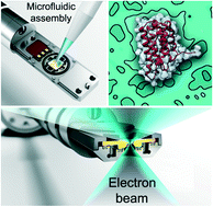
Understanding the properties of protein-based therapeutics is a common goal of biologists and physicians. Technical barriers in the direct observation of small proteins or therapeutic agents can limit our knowledge of how they function in solution and in the body. Electron microscopy (EM) imaging performed in a liquid environment permits us to peer into the active world of cells and molecules at the nanoscale. Here, we employ liquid cell EM to directly visualize a protein-based therapeutic in its native conformation and aggregate state in a time-resolved manner. In combination with quantitative analyses, information from this work contributes new molecular insights toward understanding the behaviours of immunotherapies in a solution state that mimics the human body.


 Please wait while we load your content...