Collagen peptides ameliorate intestinal epithelial barrier dysfunction in immunostimulatory Caco-2 cell monolayers via enhancing tight junctions
Abstract
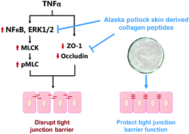
Dysfunction of the intestinal barrier plays a key role in the pathogenesis of inflammatory bowel disease (IBD) and multiple organ failure. The effect of Alaska pollock skin-derived collagen and its 3 tryptic hydrolytic fractions, HCP (6 kDa retentate), MCP (3 kDa retentate) and LCP (3 kDa permeate) on TNF-α induced barrier dysfunction was investigated in Caco-2 cell monolayers. TNF-α induced barrier dysfunction was significantly attenuated by the collagen and its peptide fractions, especially LCP, compared to TNF-α treated controls (P < 0.05). Compared to a negative control, 24 h pre-incubation with 2 mg mL−1 LCP significantly alleviated the TNF-α induced breakdown of the tight junction protein ZO-1 and occludin and inhibited MLC phosphorylation and MLCK expression. The activation of NFκB and Elk-1 was suppressed by LCP. Thus, collagen peptides may attenuate TNF-α induced barrier dysfunction of Caco-2 cells by inhibiting the NFκB and ERK1/2-mediated MLCK pathway with associated decreases in ZO-1 and occludin protein expression.


 Please wait while we load your content...