Bioceramic nanocomposite thiol-acrylate polyHIPE scaffolds for enhanced osteoblastic cell culture in 3D†
Abstract
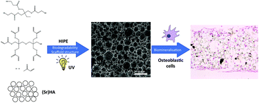
Emulsion-templated (polyHIPE) scaffolds for bone tissue engineering were produced by photopolymerisation of a mixture of trimethylolpropane tris(3-mercaptopropionate) and dipentaerythritol penta-/hexa-acrylate in the presence of hydroxyapatite (HA) or strontium-modified hydroxyapatite (SrHA) nanoparticles. Porous and permeable polyHIPE materials were produced regardless of the type or incorporation level of the bioceramic, although higher loadings resulted in a larger average pore diameter. Inclusion of HA and SrHA into the scaffolds was confirmed by EDX-SEM, FTIR and XPS and quantified by thermogravimetry. Addition of HA to polyHIPE scaffolds significantly enhanced compressive strength (148–216 kPa) without affecting compressive modulus (2.34–2.58 MPa). The resulting materials were evaluated in vitro as scaffolds for the 3D culture of MG63 osteoblastic cells vs. a commercial 3D cell culture scaffold (Alvetex®). Cells were able to migrate throughout all scaffolds, achieving a high density by the end of the culture period (21 days). The presence of HA and in particular SrHA gave greatly enhanced cell proliferation, as determined by staining of histological sections and total protein assay (Bradford). Furthermore, Von Kossa and Alizarin Red staining demonstrated significant mineralisation from inclusion of bioceramics, even at the earliest time point (day 7). Production of alkaline phosphatase (ALP), an early osteogenic marker, was used to investigate the influence of HA and SrHA on cell function. ALP levels were significantly reduced on HA- and SrHA-modified scaffolds by day 7, which agrees with the observed early onset of mineralisation in the presence of the bioceramics. The presented data support our conclusions that HA and SrHA enhance osteoblastic cell proliferation on polyHIPE scaffolds and promote early mineralisation.


 Please wait while we load your content...