Injectable silver nanosensors: in vivo dosimetry for external beam radiotherapy using positron emission tomography†
Abstract
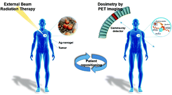
Development of safe and efficient radiotherapy routines requires quantification of the delivered absorbed dose to the cancer tissue in individual patients. In vivo dosimetry can provide accurate information about the absorbed dose delivered during treatment. In the current study, a novel silver-nanosensor formulation based on poly(vinylpyrrolidinone)-coated silver nanoparticles formulated in a gelation matrix composed of sucrose acetate isobutyrate has been developed for use as an in vivo dosimeter for external beam radiotherapy. In situ photonuclear reactions trigger the formation of radioactive 106Ag, which enables post treatment verification of the delivered dose using positron emission tomography imaging. The silver-nanosensor was investigated in a tissue equivalent thorax phantom using clinical settings and workflow for both standard fractionated radiotherapy (2 Gy) and stereotactic radiotherapy (10- and 22 Gy) in a high-energy beam setting (18 MV). The developed silver-nanosensor provided high radiopacity on the planning CT-scans sufficient for patient positioning in image-guided radiotherapy and provided dosimetric information about the absorbed dose with a 10% and 8% standard deviation for the stereotactic regimens, 10 and 22 Gy, respectively.


 Please wait while we load your content...