
Fabrication of AS1411 aptamer functionalized Gd2O3-based molecular magnetic resonance imaging (mMRI) nanoprobe for renal carcinoma cell imaging†
Abstract
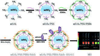
Magnetic resonance imaging (MRI) as a noninvasive diagnostic technology with high spatial resolution has been widely used in clinics. However, the relatively low sensitivity is the main shortcoming of this technology. To address this issue, we would like to develop a molecular MRI nanoprobe for the sensitive and specific MRI of renal carcinoma cells with BSA-Gd2O3 nanoparticles as MRI contrast agents, mesoporous silica nanoparticles (mSiO2 NPs) as nanocarriers and the AS1411 aptamer as a targeting molecule. To achieve this aim, BSA-Gd2O3 NPs were assembled onto mSiO2 NPs with the help of anionic polyelectrolyte, sodium polystyrene sulfonate (PSS), and cationic polyelectrolyte, poly dimethyl diallyl ammonium chloride (PDDA) layer by layer. Such successful assembly was confirmed by transmission electron microscopy (TEM), FT-IR spectroscopy, zeta-potential analysis, hydrodynamic diameter determination and gel electrophoresis. After assembly, the mSiO2/PSS/PDDA/BSA-Gd2O3 nanoprobe presented a larger longitudinal relaxivity (r1) (26.1 s−1 mM−1 Gd) than BSA-Gd2O3 NPs (11.8 s−1 mM−1 Gd) and commercially used Gd–DTPA (3.87 s−1 mM−1 Gd). Additionally, with the AS1411 aptamer as a targeting molecule, our fabricated mSiO2/PSS/PDDA/BSA-Gd2O3-AS1411 nanoprobes could recognize clear cell renal carcinoma cells (ccRCC) specifically by MRI in vitro.
 Please wait while we load your content...

