
Blue fluorogenic probes for cell plasma membranes fill the gap in multicolour imaging†
Abstract
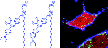
Blue fluorescent probes for cellular imaging are poorly developed and rarely used because of strong cell auto-fluorescence at these wavelengths. However, multi-colour imaging needs blue probes, such as ubiquitous nucleus markers DAPI and Hoechst, because they can be readily combined with common green and red markers based on dyes and fluorescent proteins. Cell plasma membrane is an important target for imaging, but membrane probes that absorb and emit light between 400 and 500 nm are missing. Here, using 3-methoxychromone dyes we designed two blue membrane probes exhibiting >100-fold fluorescence turn-on (fluorogenic response) on membrane binding, large Stokes shift (70–90 nm) as well as high brightness and photostability. These unique properties enabled cellular imaging at low probe concentrations (20–50 nM) with minimal background from cell auto-fluorescence and from free probe. RGB multicolour imaging was successfully realized using these probes in combination with common green and red markers. As the new probes enable high-quality imaging of cell plasma membranes in the poorly explored blue spectral region, they may become popular tools that fill the gap in multi-colour microscopy.
 Please wait while we load your content...

