Pursuing shell-isolated nanoparticle-enhanced Raman spectroscopy (SHINERS) for concomitant detection of breast lesions and microcalcifications†
Abstract
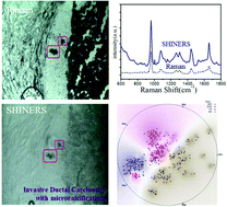
Although tissue staining followed by morphologic identification remains the gold standard for diagnosis of most cancers, such determinations relying solely on morphology are often hampered by inter- and intra-observer variability. Vibrational spectroscopic techniques, in contrast, offer objective markers for diagnoses and can afford disease detection prior to alterations in cellular and extracellular architecture by furnishing a rapid “omics”-like view of the biochemical status of the probed specimen. Here, we report a classification approach to concomitantly detect microcalcification status and local pathological state in breast tissue, featuring a combination of vibrational spectroscopy that focuses on the tumor and its microenvironment, and multivariate data analysis of spectral markers reflecting molecular expression. We employ the unprecedented sensitivity and exquisite molecular specificity offered by Au@SiO2 shell-isolated nanoparticle-enhanced Raman spectroscopy (SHINERS) to probe the presence of calcified deposits and distinguish between normal breast tissues, fibroadenoma, atypical ductal hyperplasia, ductal carcinoma in situ (DCIS), and invasive ductal carcinoma (IDC). By correlating the spectra with the corresponding histologic assessment, we developed partial least squares-discriminant analysis derived decision algorithm that provides excellent diagnostic power in the fresh frozen sections (overall accuracy of 99.4% and 93.6% using SHINs for breast lesions with and without microcalcifications, respectively). The performance of this decision algorithm is competitive with or supersedes that of analogous algorithms employing spontaneous Raman spectroscopy while enabling facile detection due to the considerably higher intensity of SHINERS. Our results pave the way for rapid tissue spectral pathology measurements using SHINERS that can offer a novel stain-free route to accurate and economical diagnoses without human interpretation.
 Please wait while we load your content...

