
Ultraporous nanofeatured PCL–PEO microfibrous scaffolds enhance cell infiltration, colonization and myofibroblastic differentiation†
Abstract
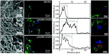
In the field of tissue engineering, integration of micro-porosity, nano-topogaphical features and weattability into one three-dimensional (3D) scaffold remains a challenge. The extracellular matrix (ECM) mimicking feature of electrospun fibers endows them wide applications in tissue engineering. However, the tight-packing of electrospun submicron fibers hinder cell infiltration and further colonization. In this study, we fabricated hydrophilic, micro-porous scaffolds with nano-topographical cues by one-step electrospinning, and investigated NIH3T3 fibroblasts cell infiltration, colonization and myofibroblastic differentiation. The hierarchical porosity enhanced cell infiltration and proliferation significantly. Besides, the nano-topography influenced the cell actin distribution and cell morphology that stimulated myofibroblastic differentiation in a drastically different manner from that of traditional solid, smooth electrospun fibers, which may hold great potential in reconstructing tissues that require strong contractile forces.
 Please wait while we load your content...

