
Quantitating membrane bleb stiffness using AFM force spectroscopy and an optical sideview setup†
Abstract
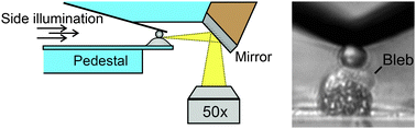
AFM-based force spectroscopy in combination with optical microscopy is a powerful tool for investigating cell mechanics and adhesion on the single cell level. However, standard setups featuring an AFM mounted on an inverted light microscope only provide a bottom view of cell and AFM cantilever but cannot visualize vertical cell shape changes, for instance occurring during motile membrane blebbing. Here, we have integrated a mirror-based sideview system to monitor cell shape changes resulting from motile bleb behavior of Xenopus cranial neural crest (CNC) cells during AFM elasticity and adhesion measurements. Using the sideview setup, we quantitatively investigate mechanical changes associated with bleb formation and compared cell elasticity values recorded during membrane bleb and non-bleb events. Bleb protrusions displayed significantly lower stiffness compared to the non-blebbing membrane in the same cell. Bleb stiffness values were comparable to values obtained from blebbistatin-treated cells, consistent with the absence of a functional actomyosin network in bleb protrusions. Furthermore, we show that membrane blebs forming within the cell–cell contact zone have a detrimental effect on cell–cell adhesion forces, suggesting that mechanical changes associated with bleb protrusions promote cell–cell detachment or prevent adhesion reinforcement. Incorporating a sideview setup into an AFM platform therefore provides a new tool to correlate changes in cell morphology with results from force spectroscopy experiments.
 Please wait while we load your content...