Tip-enhanced Raman spectroscopy of graphene-like and graphitic platelets on ultraflat gold nanoplates†
Abstract
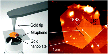
In this study, tip-enhanced Raman spectroscopy (TERS) is used to characterize graphene-like and graphitic platelets composed of a few layers of graphene. Specifically, gap-mode TERS geometry provides a larger enhancement of the local electromagnetic field at the junction formed by a gold sharp tip and a gold substrate. Graphene-like platelets are deposited onto ultra-flat thin gold nanoplates using a surfactant-assisted method. Au-coated atomic force microscopy (AFM) tips are used to probe specific substrate regions coated by the platelets. TERS spectra are collected on distinctive points on the graphene-like layers and surrounding substrate using radially or linearly polarized light, with an excitation wavelength of 632.8 nm. The position, width and intensity of G, D, and 2D Raman-active modes of graphene are discussed as a function of the incident light polarization and for distinct positions on the graphene layer. We report here on the nature of the collected TERS spectra focusing in particular on the edges of the graphene platelets.

- This article is part of the themed collection: Surface-enhanced spectroscopies

 Please wait while we load your content...